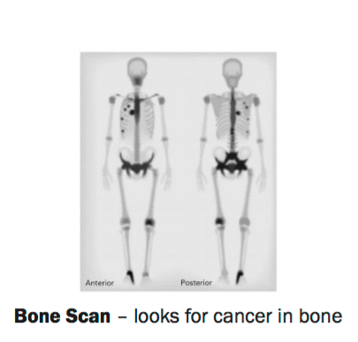
This can be due. In a normal bone scan performed 2 4 hours after radiotracer injection figure 1 some areas appear more intense compared with the rest of the skeleton. Other possible mechanisms of increased activity on bone scans include increased blood flow increased mineralisation of osteoid and interrupted sympathetic nerve supply.

In cancer diagnosis it is more usual to scan the whole body. A bone scan can look at a particular joint or bone. It is usually done in the medical physics department nuclear medicine department or x ray department of the hospital.
It is also called a radionuclide scan or a scintigram. A bone scan looks for changes or abnormalities in the bones. During a bone scan a radioactive substance called a tracer is injected into a vein in your arm.

A bone scan can often find a problem days to months earlier than a regular x ray test. A bone scan is a test that can find damage to the bones find cancer that has spread to the bones and watch problems such as infection and trauma to the bones. Tumors and cancer can cause either type.

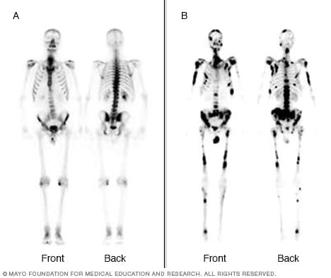
Bone loss may cause cold spots while arthritis infections or broken bones can cause hot spots. Cold spots form in areas where the tracer has not accumulated. Hot spots form when the radioactive material or tracer accumulates in areas of the bone.

Generally there are two types of spots on a bone scan. White spots in a bone scan indicate improper bone metabolism and can result from a number of conditions. Bone scans are also called scintigraphy or musculoskeletal imaging.
Bone scans are nuclear imaging tests that are used to track and diagnose various bone diseases.
Nuclear imaging bright white spots on bone scan. While bone scans are very good at finding where bone problems lie as indicated by the white spots it is often not possible to determine the cause of the problems without further tests and or a full patient medical history according to the mayo clinic 1. A bone scan that reveals white spots will often lead a doctor to order further imaging tests including a computerized tomography scan or a. With the notable exception of avascular necrosis which shows up as cold spots on a bone scan all the other above mentioned conditions show up as hot spots. Common osteoporosis hot spots to see on a bone scan include the upper thoracic spine mid back the hip joints and or the wrists.
Osteoporosis leads to fractures and bone pain. A bone scan is a nuclear imaging procedure. In nuclear imaging tiny amounts of radioactive materials tracers are injected into a vein and taken up in varying amounts at different sites in the body. Areas of the body where cells and tissues are repairing themselves most actively take up the largest amounts of tracer.
Look for hot and cold spots. Bone scans use a mildly radioactive dye to measure bone metabolism. Hot areas areas that are abnormally bright on the scan indicate that there is increased bone turnover in that area whereas cold areas on the scan abnormally dark compared with the surrounding bone indicate that there is less bone breakdown and regrowth in an area 2. Nuclear scans make pictures based on the body s chemistry like metabolism rather than on physical shapes and forms as is the case with other imaging tests.
These scans use liquid substances called radionuclide s also called tracers or radiopharmaceuticals that release low levels of radiation.

These scans use liquid substances called radionuclide s also called tracers or radiopharmaceuticals that release low levels of radiation. Nuclear scans make pictures based on the body s chemistry like metabolism rather than on physical shapes and forms as is the case with other imaging tests. Hot areas areas that are abnormally bright on the scan indicate that there is increased bone turnover in that area whereas cold areas on the scan abnormally dark compared with the surrounding bone indicate that there is less bone breakdown and regrowth in an area 2.

Bone scans use a mildly radioactive dye to measure bone metabolism. Look for hot and cold spots. Areas of the body where cells and tissues are repairing themselves most actively take up the largest amounts of tracer.
In nuclear imaging tiny amounts of radioactive materials tracers are injected into a vein and taken up in varying amounts at different sites in the body. A bone scan is a nuclear imaging procedure. Osteoporosis leads to fractures and bone pain.

Common osteoporosis hot spots to see on a bone scan include the upper thoracic spine mid back the hip joints and or the wrists. With the notable exception of avascular necrosis which shows up as cold spots on a bone scan all the other above mentioned conditions show up as hot spots. A bone scan that reveals white spots will often lead a doctor to order further imaging tests including a computerized tomography scan or a.

While bone scans are very good at finding where bone problems lie as indicated by the white spots it is often not possible to determine the cause of the problems without further tests and or a full patient medical history according to the mayo clinic 1.