
Cases with gastric dysplasia like epithelial atypia were significantly associated with a flat gross appearance a patchy distribution foveolar and gland involvement surface maturation an open nuclear chromatin pattern with prominent nucleoli retention of nuclear polarity mitoses confined to the pits lack of atypical mitoses cytoplasmic. Thus use of ck5 6 immunohistochemical staining may be helpful in the differential diagnosis as usual ductal hyperplasia is positive in a mosaic or patchwork pattern and atypical ductal hyperplasia is negative for ck5 6. Usual ductal hyperplasia and atypical ductal hyperplasia of the breast may sometimes be difficult to distinguish morphologically.

Koilocytes may have the following cellular changes. Koilocytosis or koilocytic atypia or koilocytotic atypia are terms used in histology and cytology to describe the presence of koilocytes in a specimen. A koilocyte is a squamous epithelial cell that has undergone a number of structural changes which occur as a result of infection of the cell by human papillomavirus.

The follicular cells display some degree of nuclear crowding with mild nuclear enlargement occasional oval nuclei some cells exhibiting pale chromatin and an occasional nuclear groove without evidence of nuclear inclusions papanicolaou stain original magnification 400 1000. This case was categorized as atypical primarily because of nuclear features on cytology. The related concept of dysplasia refers to an.

Atypia can be caused by an infection or irritation if diagnosed in a pap smear for example in the uterus it is more likely to be precancerous. It is used to describe atypical cells. A condition of being irregular or nonstandard is a histopathologic term for a structural abnormality in a cell i e.

Atypia from greek a typos without type. The malignancy rate for nodules that featured nuclear atypia was significantly higher at 36 8 than the rate for nodules that had only architectural atypia at 14 7 p 01. Final histology yielded 37 27 malignancies.
In total 309 thyroid nodules were diagnosed as aus flus and 137 44 were surgically excised. Nuclear atypia can be seen in reactive changes pre neoplastic changes and malignancy severe nuclear atypia is in most cases considered an indicator of malignancy. Nuclear atypia refers to abnormal appearance of cell nuclei it is a term used in cytopathology and histopathology atypical nuclei are often pleomorphic.
The lesional cells demonstrate marked nuclear atypia macronucleoli cytoplasmic keratinization and background tumor diathesis as noted there are many overlapping cytomorphologic features with hsil and differentiation may be difficult.
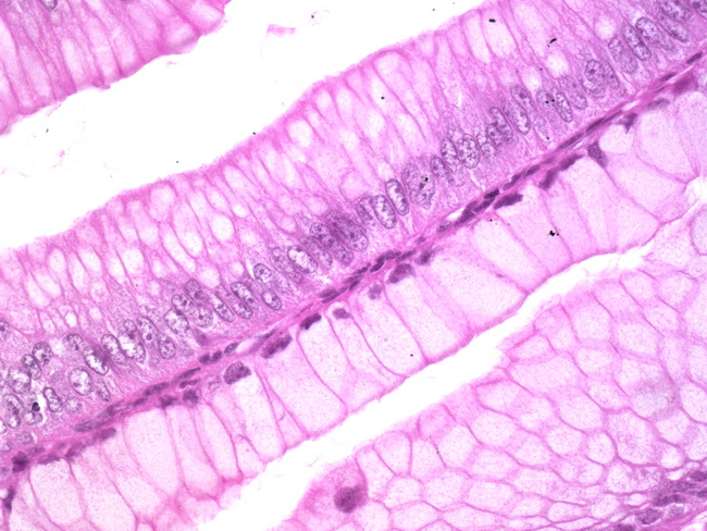
Histology nuclear atypia. Nuclear atypia also atypical nuclei is an abnormal change of the cell nucleus. Nuclear atypia is often seen in malignancy. However it may be seen in benign conditions notably inflammation also nuclear atypia in endocrine organs known as endocrine atypia is considered normal. Cancer with no appreciable or minimal nuclear atypia.
Nuclear atypia alone is generally insignificant. However with atypia loss of neurofibroma architecture high cellularity and or mitotic activity 1 50 but 3 10 high power fields the findings are worrisome for malignancy. Eric yang joel m. Palefsky in diagnostic gynecologic and obstetric pathology third edition 2018.

Palefsky in diagnostic gynecologic and obstetric pathology third edition 2018. Eric yang joel m. However with atypia loss of neurofibroma architecture high cellularity and or mitotic activity 1 50 but 3 10 high power fields the findings are worrisome for malignancy.

Nuclear atypia alone is generally insignificant. Cancer with no appreciable or minimal nuclear atypia. However it may be seen in benign conditions notably inflammation also nuclear atypia in endocrine organs known as endocrine atypia is considered normal.

Nuclear atypia is often seen in malignancy. Nuclear atypia also atypical nuclei is an abnormal change of the cell nucleus.


